Category:Images donated by Carl Zeiss Microscopy - Wikimedia Commons
The following 200 files are in this category, out of 249 total.
1869 Abbe Kondensor (7039028123).jpg
711 × 1,024; 90 KB
1877 Abbe Immersion (6892932610).jpg
715 × 1,024; 74 KB
1884 Schott Glassworks Jena (6892931538).jpg
960 × 679; 129 KB
1887 Microphotographic Apparatus by Roderich Zeiss (6892932700).jpg
1,024 × 451; 46 KB
1894 Carl Zeiss subsidiary in London (6892932398).jpg
613 × 960; 97 KB
1904 Ultraviolet Microscope (6892932882).jpg
698 × 1,024; 76 KB
1936 Pancratic Condenser (6892933212).jpg
1,024 × 733; 84 KB
1936 Phase Contrast Prototype (7039028613).jpg
718 × 1,024; 51 KB
1955 Photomicroscope Production at Winkel-Zeiss in Goettingen (7039029511).jpg
800 × 1,056; 598 KB
1957 The Winkel-Zeiss Microscope Manufacture.gif
711 × 500; 44 KB
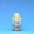
1959 Ultrafluar Objectives (7039029541).jpg
300 × 300; 13 KB
1973 Axiomat (7039029671).jpg
893 × 768; 61 KB
1986 Microscope Stands - The first 'Pyramids' (7039029805).jpg
1,024 × 755; 67 KB
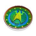
2 euro coin, confocal topography scan with ZEISS LSM 800 for Materials (27052080413).jpg
4,725 × 4,725; 1.51 MB
2013 Best of Cell Picture Show (11833473133).jpg
1,108 × 750; 345 KB
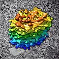
Algal Golgi body, 3D reconstruction (30466939665).jpg
2,043 × 2,043; 478 KB
August Köhler (1866-1948) (8527804902).jpg
1,262 × 1,797; 997 KB
AURIGA 60 FIB-SEM (6908574865) (2).jpg
2,952 × 2,581; 501 KB
Axio Examiner (6908552663).jpg
8,071 × 4,594; 3.83 MB
Axio Imager for Materials Microscopy (6908577871).jpg
5,368 × 3,720; 2.05 MB
Axio Imager with ApoTome.2 for Fluorescence Optical Sectioning (9296865359).jpg
7,495 × 4,823; 2.85 MB
Axio Imager with Colibri.2 LED Lightsource for Fluorescence Illumination (9299647318).jpg
2,976 × 3,856; 1.03 MB
Axio Observer with ApoTome.2 (6908584785).jpg
9,449 × 4,868; 2.88 MB
Axio Vert.A1 (6908550157).jpg
7,171 × 4,487; 4.14 MB
Axio Vert.A1 (6908578857).jpg
4,992 × 6,668; 1.16 MB
Axio Vert.A1 for Materials Microscopy (6908554003).jpg
4,724 × 2,858; 1.52 MB
Axio Zoom.V16 with ApoTome.2 (6908563695).jpg
8,585 × 4,988; 2.51 MB
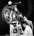
Axiomat with BMW motor cycle on top (6895200642).jpg
1,879 × 1,966; 449 KB
Binocular compound microscope, Carl Zeiss Jena, 1914 (6779276516).jpg
2,400 × 3,600; 3.25 MB
Bringing Biology Into Focus (15718801672).jpg
1,280 × 847; 968 KB
C. elegans, model organism in life sciences (28703152561).jpg
2,560 × 1,920; 369 KB
Carl Zeiss (6909161791).jpg
2,185 × 2,991; 568 KB
Carl Zeiss Jena (7039030311).jpg
1,000 × 1,000; 191 KB
Carl Zeiss production in Jena, ca. 1890 (6892931876) (cropped).jpg
782 × 561; 120 KB
Carl Zeiss production in Jena, ca. 1890 (6892931876).jpg
796 × 566; 116 KB
Carlos Chagas.png
659 × 772; 361 KB
CeBER Outreach 2014 (15420289914).jpg
2,835 × 1,862; 1.56 MB
CeBER Outreach 2014 (15856511009).jpg
2,835 × 1,894; 1.43 MB
CeBER Outreach 2014 (15856797127).jpg
2,835 × 1,874; 1.15 MB
CeBER Outreach 2014 (16040608261).jpg
2,835 × 1,900; 1.53 MB
Cell Observer (6908566097).jpg
4,724 × 1,896; 812 KB
Cell Observer SD (6908586101).jpg
4,096 × 2,372; 862 KB
Correlative 3D microscopy of plasmodesmata inside root nodules (25236962785).jpg
1,291 × 764; 193 KB
Correlative Microscopy System Shuttle & Find - MERLIN and Axio Observer (6908581125).jpg
10,361 × 5,852; 3.56 MB
Correlative Microscopy with ZEISS Shuttle&Find - MERLIN and LSM 800 (16292616486).jpg
5,156 × 2,362; 976 KB
Cross section of printed circuit board (25140841400).jpg
2,809 × 2,105; 436 KB
Cultured Rat Hippocampal Neuron (24327909026).jpg
1,918 × 1,219; 784 KB
Diffractive optics (24564443291).jpg
800 × 600; 386 KB
Early human embryos (27872285595).jpg
1,300 × 1,030; 204 KB
Ernst Abbe (6892931486).jpg
583 × 960; 42 KB
Ernst Abbe (6909162215).jpg
1,891 × 2,607; 524 KB
EVO HD 15 (6908552901).jpg
1,540 × 1,540; 201 KB
Fluorescence Dynamics and Photomanipulation (10690270154).jpg
5,903 × 8,259; 3.11 MB
Fluorescence Microscope ZEISS UNIVERSAL (ca. 1965) (6834822839).jpg
600 × 443; 39 KB
Fluorescence microscopy with ZEISS Axiocam (cropped).png
985 × 763; 353 KB
Fluorescence microscopy with ZEISS Axiocam.png
1,500 × 767; 588 KB
Fluorescent Dyes and Proteins (10690288626).jpg
5,903 × 8,259; 2.02 MB
Grave of Carl Zeiss in Jena, Germany (7039027209).jpg
641 × 960; 217 KB
Hans Boegehold (7039028871).jpg
679 × 977; 86 KB
Hazelnut (male flower), overlay of 7 channel autofluorescence microscopy (30458886372).jpg
8,396 × 5,059; 4.36 MB
How big is 1 micrometer? (10690468113) (2).jpg
5,903 × 8,259; 3.11 MB
How big is 1 micrometer? (10690468113).jpg
5,506 × 6,886; 3.4 MB
Human embryos (four-cell stage) (27771482412).jpg
650 × 496; 58 KB
Imaging Life with Fluorescent Proteins (10690274384) (2).jpg
5,903 × 8,259; 3.8 MB
Imaging Life with Fluorescent Proteins (10690274384).jpg
5,531 × 6,876; 3.32 MB
Indian Muntjac fibroblast cells (23725924864).jpg
1,924 × 1,218; 718 KB
Indian Muntjac fibroblast cells (24271618921).jpg
1,923 × 1,210; 684 KB
Indian Muntjac fibroblast cells (24327908636).jpg
1,920 × 1,217; 1,007 KB
Kidney section, fluorescence microscopy (30575642655).jpg
12,182 × 7,147; 6.17 MB
Köhler Illumination with the Inverted Microscope (15174751101).jpg
3,000 × 4,200; 638 KB
Köhler Illumination with the Upright Microscope (15177755065).jpg
3,000 × 4,200; 672 KB
Labscope for Windows (30192942961).jpg
8,688 × 5,792; 4.76 MB
Laser structured surface (24019830053).jpg
800 × 600; 335 KB
Laser TIRF 3 (6908564909).jpg
5,906 × 4,000; 1.32 MB
Laser TIRF 3 (6908574263).jpg
5,906 × 4,000; 1.38 MB
Life Science Imaging with ZEISS Scanning Electron Microscopes (10690272504) (2).jpg
5,903 × 8,259; 4.3 MB
Life Science Imaging with ZEISS Scanning Electron Microscopes (10690272504).jpg
5,614 × 6,876; 3.9 MB
LSM 7 MP (6908576283).jpg
7,087 × 4,495; 2.16 MB
LSM 700 (6908567127).jpg
7,087 × 3,366; 1.49 MB
LSM 780 NLO (6908556701).jpg
7,087 × 3,854; 1.45 MB
Material Analysis with ZEISS Scanning Electron Microscopes (10690229705) (2).jpg
5,903 × 8,260; 3.54 MB
Material Analysis with ZEISS Scanning Electron Microscopes (10690229705).jpg
5,624 × 6,887; 3.18 MB
Mausoleum of Ernst Abbe in Jena, Germany (6892932920).jpg
450 × 600; 62 KB
Metallographic system based on Martens design, ca. 1904 (6892932760).jpg
1,024 × 717; 92 KB
Michel-Lévy interference colour chart (21257606712).jpg
11,586 × 8,177; 3.25 MB
Microfluidic channel, confocal 3D view (26183999723).jpg
4,488 × 3,069; 2.34 MB
Milled aluminum surface (24278900649).jpg
800 × 600; 260 KB
Mitotic LLC-PK1 cells, fluorescence microscopy (23700644352).jpg
1,800 × 1,200; 1.6 MB
Mobile phone camera lens module, 3D X-ray microscopy (30033111412).jpg
8,592 × 7,823; 7.56 MB
Mouse Intestine (23727287703).jpg
1,915 × 1,217; 1,010 KB
Mouse Kidney (23725924684).jpg
1,916 × 1,210; 993 KB
Mouse tissue, stained histology preparation (23180949464).jpg
4,248 × 2,832; 2.19 MB
Mouse tissue, stained histology preparation (23782972906).jpg
4,248 × 2,832; 1.97 MB
Multi-discussion system for ZEISS Axio Lab.A1 (10474362435).jpg
5,616 × 3,744; 2.28 MB
Nanofluidics channels (33411553986).jpg
6,144 × 4,608; 10.09 MB
Nanosize 3D model of a vintage ZEISS microscope, by Nanoscribe (29829841445).jpg
1,457 × 970; 206 KB
Nanoworlds (29746468381).jpg
2,048 × 1,536; 1.18 MB
New ZEISS Axio Observer Family for Metallurgy and Materials Imaging (21673571488).jpg
5,616 × 3,635; 1.51 MB
Oocyte with Zona pellucida (27771482282).jpg
1,300 × 1,030; 153 KB
Opening of the ZEISS Forum and Museum of Optics (14553532890).jpg
2,738 × 1,825; 1.34 MB
Opening of the ZEISS Forum and Museum of Optics (14553533450).jpg
2,738 × 1,825; 2.4 MB
Opening of the ZEISS Forum and Museum of Optics (14553539758).jpg
2,738 × 1,825; 1.16 MB
Opening of the ZEISS Forum and Museum of Optics (14553540838).jpg
2,828 × 1,767; 1.28 MB
Opening of the ZEISS Forum and Museum of Optics (14553542688).jpg
2,738 × 1,825; 1.85 MB
Opening of the ZEISS Forum and Museum of Optics (14553543198).jpg
2,696 × 1,854; 1.36 MB
Opening of the ZEISS Forum and Museum of Optics (14717192316).jpg
2,738 × 1,825; 1.13 MB
Opening of the ZEISS Forum and Museum of Optics (14737026061).jpg
2,738 × 1,825; 1.22 MB
Opening of the ZEISS Forum and Museum of Optics (14739906372).jpg
2,738 × 1,825; 1.29 MB
Opening of the ZEISS Forum and Museum of Optics (14740195125).jpg
2,181 × 1,198; 858 KB
Opening of the ZEISS Forum and Museum of Optics (14740195495).jpg
2,738 × 1,825; 1.26 MB
Opening of the ZEISS Forum and Museum of Optics (14760055523).jpg
2,433 × 2,055; 1.32 MB
Opening of the ZEISS Forum and Museum of Optics (14760057043).jpg
2,738 × 1,825; 1.5 MB
ORION NanoFab - Helium Ion Microscope (8410606251).jpg
3,107 × 3,388; 898 KB
Otto Schott (7039028041).jpg
612 × 1,024; 64 KB
PALM CombiSystem by ZEISS Microscopy (7948945926).jpg
945 × 494; 78 KB
Paper surface with indent from pen, confocal 3D view (26515185690).jpg
2,481 × 1,653; 938 KB
ParticleSCAN VP (6908567401).jpg
1,540 × 1,540; 264 KB
Primo Star by ZEISS - Your compact microscope for education and routine (8864950302).jpg
5,470 × 2,735; 1.28 MB
Printed graphite circuit on substrate, confocal 3D view (26720581401).jpg
2,244 × 1,917; 706 KB
Rat primary cortical neuron culture, 3D reconstruction (30614936992).jpg
1,643 × 1,052; 629 KB
Rat primary cortical neuron culture, deconvolved z-stack overlay (30614937102).jpg
2,752 × 2,208; 1.86 MB
Robert Koch (6909161361).jpg
1,360 × 1,950; 916 KB
Rudolf Winkel (6892933560).jpg
306 × 420; 47 KB
Santiago Ramón y Cajal (8691434507).jpg
939 × 1,059; 57 KB
Santiago Ramón y Cajal (8691434605).jpg
2,278 × 2,068; 724 KB
Science Poster- The Scanning Electron Microscope (23377045739).jpg
3,672 × 5,556; 1.68 MB
Scratch on glass surface, 3D topography (26711616455).jpg
2,363 × 1,463; 321 KB
SIGMA (6908562113).jpg
1,536 × 1,651; 286 KB
SK8-18-2 human derived cells, fluorescence microscopy (29942101073).jpg
2,758 × 2,214; 450 KB
Spider, false-coloured scanning electron micrograph (27813345024).jpg
1,024 × 768; 1.06 MB
Stand I from 1891, Optical Workshop Carl Zeiss Jena (28918277463).jpg
1,500 × 2,862; 2.75 MB
Stereoscan MK1 (19515061054).jpg
3,910 × 2,827; 661 KB
SUPRA 55 (6908563989).jpg
1,644 × 1,632; 350 KB
Supra VP (6908585171).jpg
2,157 × 2,024; 461 KB
The Abbe Formula (6892931394).jpg
717 × 960; 36 KB
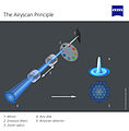
The Airyscan Principle (14657210018).jpg
2,117 × 2,154; 570 KB
The Carl Zeiss Optical Workshop 1896 in Jena, Germany (6892931982) (2).jpg
2,083 × 1,483; 4.24 MB
The Carl Zeiss Optical Workshop 1896 in Jena, Germany (6892931982).jpg
960 × 744; 163 KB
ULTRA (6908561771).jpg
2,662 × 2,240; 530 KB
ZEISS and Cell Press present the Cell Picture Show (14989291035).jpg
1,800 × 1,500; 2.67 MB
ZEISS at Neuroscience 2016 (30496671074).jpg
4,597 × 2,969; 6.74 MB
ZEISS at Neuroscience 2016 (30511114503).jpg
4,275 × 2,701; 6.69 MB
ZEISS at Neuroscience 2016 (30513797794).jpg
4,908 × 3,272; 12.11 MB
ZEISS at Neuroscience 2016 (30950326290).jpg
5,121 × 3,414; 6.69 MB
ZEISS at Neuroscience 2016 (30950369170).jpg
4,306 × 2,930; 5.64 MB
ZEISS at Neuroscience 2016 (30950371360).jpg
4,874 × 3,130; 6.8 MB
ZEISS at Neuroscience 2016 (30950375160).jpg
4,240 × 2,832; 3.06 MB
ZEISS at Neuroscience 2016 (30967339960).jpg
5,184 × 3,456; 10.39 MB
ZEISS at Neuroscience 2016 (31174715202).jpg
5,331 × 3,393; 10.04 MB
ZEISS at Neuroscience 2016 (31203824801).jpg
4,597 × 2,925; 7.4 MB
ZEISS at Neuroscience 2016 (31318646845).jpg
4,730 × 2,442; 5.99 MB
ZEISS at Neuroscience 2016 (31318696255).jpg
5,472 × 3,648; 8.74 MB
ZEISS AURIGA and Crossbeam FIB-SEM Technology and Applications (10690235245).jpg
5,903 × 8,260; 3.09 MB
ZEISS Autocorr Microscope Objectives (30539466086).jpg
7,554 × 5,078; 2.25 MB
ZEISS Autocorr objectives for Celldiscoverer 7 (30099434184).jpg
5,616 × 3,744; 1.72 MB
ZEISS Axio Imager.A2 (11234755945).jpg
5,616 × 3,744; 1.65 MB
ZEISS Axio Observer 3 materials (21861533455).jpg
5,510 × 5,792; 1.9 MB
ZEISS Axio Observer 5 materials (21871137271).jpg
5,371 × 5,406; 2.02 MB
ZEISS Axio Observer 7 materials (21673475730).jpg
6,675 × 5,456; 2.18 MB
ZEISS Axio Observer for Life Sciences (30458875472).jpg
8,024 × 5,382; 1.15 MB
ZEISS Axio Observer.A1 (11234767744).jpg
5,616 × 3,744; 1.45 MB
ZEISS Axio Scan.Z1 (8407041905).jpg
6,359 × 3,744; 1.4 MB
ZEISS Axio Zoom.V16 (10943206726).jpg
2,362 × 1,244; 340 KB
ZEISS Axiocam 512 color (23809080335).jpg
8,688 × 5,792; 3.46 MB
ZEISS Axiocam 702 mono (23441118979).jpg
8,688 × 5,792; 4.43 MB
ZEISS Axiocam 702 mono (23726632841).jpg
8,688 × 5,792; 4.97 MB
ZEISS C Epiplan-APOCHROMAT Objectives (24538265432).jpg
5,616 × 3,744; 1.35 MB
ZEISS Celldiscoverer 7 (30099434844).jpg
4,368 × 3,744; 898 KB
ZEISS Celldiscoverer 7 (30614937742).jpg
8,011 × 3,744; 1.63 MB
ZEISS Colibri 7 (30487876501).jpg
8,688 × 5,792; 2.03 MB
ZEISS Crossbeam (9662745943).jpg
8,858 × 4,992; 3.22 MB
ZEISS Day of Microscopy 2015 (16178429514).jpg
2,738 × 1,825; 444 KB
ZEISS Day of Microscopy 2015 (16180847243).jpg
2,738 × 1,825; 383 KB
ZEISS Day of Microscopy 2015 (16180847623).jpg
2,738 × 1,825; 347 KB
ZEISS Day of Microscopy 2015 (16593471777).jpg
2,000 × 1,492; 391 KB
ZEISS Day of Microscopy 2015 (16593471947).jpg
2,000 × 1,331; 633 KB
ZEISS Day of Microscopy 2015 (16593472667).jpg
2,738 × 1,825; 635 KB


