Category:Media from PeerJ - Wikimedia Commons
 Article Images
The following 200 files are in this category, out of 427 total.
A new species of Pseudopaludicola (Anura, Leiuperinae) from Espírito Santo, Brazil.pdf
1,275 × 1,650, 25 pages; 18.82 MB
Advertisement call of Leptodactylus apepyta.ogg
26 s; 294 KB
Advertisement call of Leptodactylus mystacinus.wav
22 s; 1.83 MB
Advertisement call of Phyllodytes magnus-2.WAV
47 s; 3.94 MB
Advertisement call of Phyllodytes magnus-3.wav
3 min 33 s; 17.92 MB
Advertisement call of Phyllodytes magnus-4.wav
3 min 48 s; 19.14 MB
Advertisement call of Phyllodytes magnus.png
1,980 × 1,289; 876 KB
Advertisement call of Pseudopaludicola restinga.png
848 × 855; 69 KB
Advertisement vocalizations of Mercurana myristicapalustris.png
2,397 × 1,952; 1.28 MB
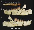
Akhnatenavus mandible Borths et al. 2016.png
2,800 × 2,477; 4.34 MB
Akhnatenavus maxilla Borths et al. 2016.png
3,242 × 1,743; 3.54 MB
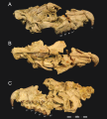
Akhnatenavus skull Borths et al. 2016.png
3,242 × 3,623; 7.87 MB
Akhnatenavus teeth Borths et al. 2016.png
3,219 × 1,897; 3.05 MB
Allosaurus "Big Al II".jpg
5,194 × 1,942; 1.04 MB
An integrative taxonomic analysis reveals a new species of lotic Hynobius salamander from Japan.pdf
1,275 × 1,650, 40 pages; 39.23 MB
Analyzing-machupo-virus-receptor-binding-by-molecular-dynamics-simulations-peerj-02-266-s001.ogv
12 s, 1,311 × 1,019; 16.36 MB
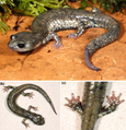
Aneides iecanus.png
1,445 × 1,187; 3.16 MB
Aneides niger from the campus of UC Santa Cruz.png
2,016 × 1,348; 5.41 MB
Aneides niger.png
993 × 655; 1.22 MB
Anhanguerid skeleton.png
2,832 × 1,861; 2.62 MB
Antibacterial properties of antimicrobial peptides (AMP) - Fig-1-2x.jpg
1,070 × 1,200; 185 KB
Asian-elephants-(Elephas-maximus)-reassure-others-in-distress-peerj-02-278-s001.ogv
51 s, 320 × 240; 5.39 MB
Astrobatrachus kurichiyana in live.png
2,437 × 3,531; 16.07 MB
Astrobatrachus kurichiyana.png
2,437 × 1,500; 5.68 MB
Atopos and Plectostoma concinnum.png
506 × 442; 441 KB
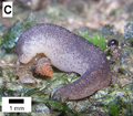
Atopos.png
477 × 293; 197 KB
Banhxeochelys carapax collage - Garbin et al 2019.png
5,261 × 3,697; 18.76 MB
Behavior and feeding of Endeavouria septemlineata.png
2,807 × 1,961; 10.63 MB
Birkamys dentary Sallam et al 2016.png
4,252 × 3,040; 10.9 MB
Birkamys holotype Sallam et al 2016.png
4,252 × 4,982; 19.46 MB
Birkamys lower dentition Sallam et al 2016.png
4,252 × 3,237; 9.02 MB
Birkamys upper dentition Sallam et al 2016.png
4,252 × 3,247; 10.93 MB
Brachycephalus auroguttatus.png
1,786 × 1,231; 2.95 MB
Brachycephalus boticario.png
1,786 × 1,204; 3.21 MB
Brachycephalus brunneus.png
2,113 × 1,401; 5.76 MB
Brachycephalus coloratus.png
1,772 × 1,181; 3.19 MB
Brachycephalus curupira 2.png
2,105 × 1,393; 6.55 MB
Brachycephalus curupira.png
1,772 × 1,181; 3.27 MB
Brachycephalus fuscolineatus.png
1,729 × 1,154; 3.13 MB
Brachycephalus izecksohni.png
2,119 × 1,369; 5.44 MB
Brachycephalus leopardus 2.png
2,089 × 1,369; 5.16 MB
Brachycephalus leopardus amplexus.png
1,786 × 1,204; 2.97 MB
Brachycephalus leopardus.png
1,822 × 1,204; 3.01 MB
Brachycephalus mariaeterezae.png
1,844 × 1,204; 3.34 MB
Brachycephalus olivaceus.png
1,844 × 1,231; 3.41 MB
Brachycephalus quiririensis dorsal color variation.png
1,668 × 2,394; 5.1 MB
Brachycephalus quiririensis holotype.png
2,883 × 2,883; 1.63 MB
Brachycephalus quiririensis.png
1,772 × 1,181; 2.61 MB
Brachycephalus verrucosus.png
1,786 × 1,204; 3.47 MB
Brancasaurus map.jpg
1,319 × 1,013; 202 KB
Brancasaurus-15.jpg
1,200 × 637; 243 KB
Breaching True's beaked whales.png
1,831 × 900; 2.73 MB
Breaching True´s beaked whales.jpg
1,831 × 900; 1.05 MB
Brychotherium Borths et al 2016.png
3,599 × 1,940; 3.96 MB
Brychotherium dentaries Borths et al 2016.png
3,661 × 3,881; 5.01 MB
Brychotherium dentary Holotype Borths et al 2016.png
3,242 × 2,042; 4.07 MB
Brychotherium rostrum Borths et al 2016.png
3,242 × 2,005; 4.53 MB
Brychotherium rostrum2 Borths et al 2016.png
3,212 × 2,114; 2.62 MB
Cape presentation display of superb birds-of-paradise.jpg
1,200 × 448; 153 KB
Centrosaurus skulls.png
2,664 × 3,740; 5.27 MB
Choerophryne alpestris.png
729 × 481; 796 KB
Choerophryne proboscidea.png
740 × 490; 731 KB
Choerophryne sp. 2.png
732 × 484; 481 KB
Choerophryne sp.png
743 × 484; 480 KB
Chromosome features of Pseudopaludicola restinga.png
12,323 × 4,692; 4.22 MB
Clea nigricans radula.png
1,532 × 746; 424 KB
Color patterns of Aneides flavipunctatus from the Longvale & Laytonville, CA.png
3,000 × 2,008; 10.65 MB
Color patterns of Aneides flavipunctatus.png
2,000 × 2,248; 9.74 MB
Comments-and-corrections-on-3D-modeling-studies-of-locomotor-muscle-moment-arms-in-archosaurs-peerj-03-1272-s002.ogv
11 s, 1,248 × 1,080; 1.75 MB
Comments-and-corrections-on-3D-modeling-studies-of-locomotor-muscle-moment-arms-in-archosaurs-peerj-03-1272-s003.ogv
14 s, 1,248 × 1,080; 2.57 MB
Comments-and-corrections-on-3D-modeling-studies-of-locomotor-muscle-moment-arms-in-archosaurs-peerj-03-1272-s004.ogv
14 s, 1,248 × 1,080; 2.23 MB
Comments-and-corrections-on-3D-modeling-studies-of-locomotor-muscle-moment-arms-in-archosaurs-peerj-03-1272-s005.ogv
17 s, 1,248 × 1,080; 3.34 MB
Comments-and-corrections-on-3D-modeling-studies-of-locomotor-muscle-moment-arms-in-archosaurs-peerj-03-1272-s006.ogv
14 s, 1,248 × 1,080; 2.84 MB
Contrasting color in Aneides flavipunctatus complex.png
2,659 × 2,390; 3.6 MB
Courtship and amplexus in Mercurana myristicapalustris.png
3,497 × 2,497; 15.01 MB
Craniofacial-ontogeny-in-Centrosaurus-apertus-peerj-02-252-g001.jpg
776 × 587; 108 KB
Craniofacial-ontogeny-in-Centrosaurus-apertus-peerj-02-252-g002.jpg
765 × 856; 105 KB
Craniofacial-ontogeny-in-Centrosaurus-apertus-peerj-02-252-g003.jpg
765 × 846; 104 KB
Craniofacial-ontogeny-in-Centrosaurus-apertus-peerj-02-252-g004.jpg
743 × 326; 46 KB
Craniofacial-ontogeny-in-Centrosaurus-apertus-peerj-02-252-g005.jpg
744 × 316; 44 KB
Craniofacial-ontogeny-in-Centrosaurus-apertus-peerj-02-252-g007.jpg
787 × 1,121; 143 KB
Craniofacial-ontogeny-in-Centrosaurus-apertus-peerj-02-252-g008.jpg
645 × 1,496; 144 KB
Craniofacial-ontogeny-in-Centrosaurus-apertus-peerj-02-252-g009.jpg
703 × 323; 50 KB
Diegoaelurus Canine and molar morphology - Zack et al 2022.png
2,000 × 2,455; 4.54 MB
Diegoaelurus mandible - Zack et al 2022.png
2,000 × 1,740; 2.07 MB
Distribution map of Hynobius kimurae, H. boulengeri & H. fossigenus.png
2,437 × 1,541; 1.13 MB
Distribution map of Pseudopaludicola restinga.png
901 × 900; 206 KB
Distribution of mass and interocular distance in Enteroctopus dofleini.png
2,666 × 1,852; 335 KB
Distribution of total length, mantle length and mass in Architeuthis dux.png
2,666 × 1,924; 258 KB
DNHM D2945 6 interpretive.png
1,242 × 2,010; 650 KB
DNHM D2945 6 skull.png
1,193 × 628; 1.41 MB
Dorsal and ventral views of male Maguimithrax spinosissimus, Florida.png
4,386 × 1,861; 5.69 MB
Dorsal view of Astrobatrachus kurichiyana.png
1,197 × 870; 1.84 MB
Dorsolateral view of Nyctibatrachus manalari.png
1,005 × 455; 790 KB
Dorsolateral view of Nyctibatrachus pulivijayani.png
994 × 443; 859 KB
Ear-tagged and radio-collared Macroscelides micus.png
692 × 461; 483 KB
Egg sacs and larval stage of Hynobius fossigenus.png
2,437 × 3,153; 4.29 MB
Ejection and retraction of defense apparatus acontia in sea anemone Exaiptasia pallida.webm
2 min 24 s, 853 × 480; 11.77 MB
Elaphe urartica color and pattern variation.png
2,250 × 2,288; 10.06 MB
Elasmosaurus skeletal.png
3,750 × 521; 698 KB
Electroneuria ronwoodi holotype Fig5 A.jpg
579 × 387; 287 KB
Electroneuria ronwoodi holotype Fig5 B.jpg
578 × 385; 280 KB
Electroneuria ronwoodi holotype Fig5 C.jpg
577 × 387; 249 KB
Electroneuria ronwoodi holotype Fig5 D.jpg
579 × 386; 247 KB
Electroneuria ronwoodi holotype Fig5 E.jpg
576 × 385; 209 KB
Electroneuria ronwoodi holotype Fig5 F.jpg
580 × 386; 239 KB
Electroneuria ronwoodi holotype Fig5.jpg
1,188 × 1,200; 465 KB
Endeavouria septemlineata aggregation.png
1,439 × 1,033; 3.1 MB
Endeavouria septemlineata feeding on Bradybaena similaris.png
1,256 × 1,030; 2.08 MB
Endeavouria septemlineata feeding on Rhinocricus millipede.png
1,007 × 654; 1.21 MB
Epiperipatus puri sp. nov., a new velvet worm from Atlantic Forest in Southeastern Brazil (Onychophora, Peripatidae).pdf
1,275 × 1,650, 16 pages; 21.23 MB
Fig-2-full - Haptoral and male genital sclerotized structures from Cichlidogyrus spp.png
1,855 × 2,093; 2.72 MB
First underwater video of True's beaked whales underwater.ogv
46 s, 1,920 × 1,080; 15.79 MB
Foot Drumming Somali Sengi at Assamo Djibouti - Steven.Heritage et al 2020.webm
26 s, 1,536 × 1,152; 67.64 MB
Fossil specimen (DNHM D2945 6) of Hongshanornis longicresta.jpg
1,242 × 2,004; 1.78 MB
Fukomys clades Faulkes 2017.png
2,399 × 2,991; 313 KB
Fukomys map Faulkes 2017.png
2,472 × 3,032; 10.12 MB
Fukomys molecular clock Faulkes 2017.png
2,582 × 2,675; 497 KB
Fêmea parátipo de Epiperipatus puri.jpg
1,234 × 1,757; 438 KB
Galegeeska habitat - Heritage et al 2020.png
1,619 × 1,943; 6.82 MB
Galegeeska revoili - Heritage et al 2020.png
1,619 × 1,295; 3.29 MB
Galegeeska revoili-b - Heritage et al 2020.png
1,619 × 1,295; 3.4 MB
Geckolepis megalepis crop.png
2,802 × 2,020; 6.25 MB
Geckolepis megalepis.png
4,422 × 3,963; 24.98 MB
Geographic distribution of Leptodactylus apepyta and L. mystacinus.png
3,230 × 2,687; 4.22 MB
Geographic distribution of Phyllodytes magnus.png
2,000 × 1,545; 4.74 MB
Glossotherium carnivore marks - Chichkoyan etal 2017.png
4,408 × 3,712; 15.28 MB
Goniotarbus angulatus holotype fossil A-E.jpg
891 × 1,180; 596 KB
Goniotarbus angulatus holotype fossil dorsal ventral.jpg
886 × 552; 281 KB
Habit and Habitat of Mercurana myristicapalustris.png
3,500 × 2,973; 17.45 MB
Habitats at Kingman Reef - Peerj-81-fig-2A.png
1,329 × 831; 1.92 MB
Habitats at Kingman Reef - Peerj-81-fig-2B.png
1,266 × 882; 2.38 MB
Habitats at Kingman Reef - Peerj-81-fig-2C.png
1,274 × 881; 2.35 MB
Habitats at Kingman Reef - Peerj-81-fig-2D.png
1,318 × 815; 2.48 MB
Habitats at Kingman Reef - Peerj-81-fig-2E.png
1,318 × 830; 2.04 MB
Habitats at Kingman Reef - Peerj-81-fig-2F.png
1,331 × 818; 1.63 MB
Habitats at Kingman Reef - Peerj-81-fig-2G.png
1,264 × 888; 2.32 MB
Habitats at Kingman Reef - Peerj-81-fig-2H.png
1,258 × 880; 2.07 MB
Habitats of Nidirana leishanensis in Leishan County.png
2,592 × 1,966; 9.14 MB
Heyuannia skeletal.png
2,500 × 1,400; 759 KB
High-resolution CT scan of paratype of Brachycephalus curupira.png
7,088 × 5,005; 12.06 MB
Holmesina skull - Gaudin et al 2017.png
1,867 × 1,365; 3.68 MB
Holmesina UF 191448 - Gaudin et al 2017.png
861 × 1,364; 1.08 MB
Holmesina UF 248500 - Gaudin et al 2017.png
888 × 1,362; 1.15 MB
Holotype and paratype specimens of Micryletta aishani.png
1,896 × 2,334; 6.83 MB
Holotype Fukomys hanangensis Faulkes 2017.jpg
1,453 × 1,355; 236 KB
Holotype Fukomys livingstoni Faulkes 2017.jpg
1,391 × 1,273; 236 KB
Holotype of Aneides klamathensis (cropped).png
2,000 × 1,282; 4.41 MB
Holotype of Aneides klamathensis.png
2,000 × 2,078; 8.44 MB
Holotype of Brachycephalus coloratus.png
1,635 × 1,646; 1.83 MB
Holotype of Brachycephalus curupira.png
1,584 × 1,654; 1.9 MB
Holotype of Diegoaelurus vanvalkenburghae.png
4,768 × 5,907; 20.7 MB
Holotype of Hynobius fossigenus.png
2,437 × 3,444; 5.67 MB
Holotype of Leptodactylus apepyta in preservative.png
3,230 × 3,199; 5.82 MB
Holotype of Leptodactylus apepyta.png
3,230 × 1,363; 5.78 MB
Holotype of Micryletta aishani.png
950 × 613; 897 KB
Holotype of Nidirana leishanensis in preservative.png
4,454 × 3,628; 7.12 MB
Holotype of Nyctibatrachus pulivijayani (cropped).png
5,200 × 3,773; 9.76 MB
Holotype of Nyctibatrachus pulivijayani.png
5,200 × 6,125; 30.28 MB
Holotype of Phyllodytes magnus-1.png
2,000 × 1,116; 1.63 MB
Holotype of Phyllodytes magnus-2.png
2,000 × 2,011; 3.25 MB
Holotype of Pseudopaludicola restinga in preservative 2.png
4,621 × 1,528; 6.68 MB
Holotype of Pseudopaludicola restinga in preservative.png
3,426 × 2,022; 5.7 MB
Holotypes Fukomys livingstoni and Fukomys hanangensis Faulkes 2017.png
1,531 × 2,773; 6.07 MB
Hongshanornis scale.png
1,158 × 1,185; 42 KB
Hongshanornis wingspan.png
983 × 576; 36 KB
Hynobius boulengeri.jpg
644 × 347; 109 KB
Hynobius fossigenus.png
1,876 × 1,210; 3.45 MB
Hyoid and larynx of Pseudopaludicola restinga.png
4,899 × 4,454; 9.13 MB
Hypoxia-inducible-C-to-U-coding-RNA-editing-downregulates-SDHB-in-monocytes-peerj-01-152-s008.ogv
25 s, 720 × 480; 6.62 MB
Illustration of Astrobatrachus kurichiyana.png
2,437 × 3,135; 5.98 MB
Intraspecific variation observed in Leptodactylus apepyta.png
3,240 × 2,921; 16.16 MB
Intraspecific variation observed in Leptodactylus mystacinus.png
3,240 × 2,203; 11.62 MB
Invictarx zephyri dorsal vertebrae PeerJ e5435 fig 5.png
2,428 × 2,314; 6.8 MB
Invictarx zephyri holotype PeerJ e5435 fig 3.png
2,214 × 2,593; 6.77 MB
Invictarx zephyri holotype PeerJ e5435 fig 4.png
2,428 × 1,902; 4.27 MB
Invictarx zephyri limb elements PeerJ e5435 fig 6.png
2,428 × 2,207; 5.47 MB
Karyotype of Phyllodytes magnus.png
3,732 × 650; 264 KB
Lapisperla keithrichardsi holotype Fig3 A.jpg
585 × 438; 296 KB
Lapisperla keithrichardsi holotype Fig3 B.jpg
586 × 437; 304 KB
Lapisperla keithrichardsi holotype Fig3 C.jpg
585 × 437; 397 KB
Lapisperla keithrichardsi holotype Fig3 D.jpg
585 × 439; 177 KB
Lapisperla keithrichardsi holotype Fig3.jpg
1,200 × 906; 370 KB
Largusoperla billwymani holotype fig9 A.jpg
583 × 392; 292 KB
Largusoperla billwymani holotype fig9 B.jpg
581 × 394; 312 KB
Largusoperla billwymani holotype fig9 C.jpg
582 × 437; 273 KB
Largusoperla billwymani holotype fig9 D.jpg
584 × 214; 173 KB
Largusoperla billwymani holotype fig9 E.jpg
584 × 209; 138 KB


